Lecture Six: Methods of Assessing Fetal Status


NURS 2208
T. Dennis RNC, MSN
Objectives
- Discuss the use of ultrasound in pregnancy
- Discuss methods of antenatal fetal surveillance
- Identify antenatal surveillance indicators
- Compare NST, CST and BPP
- Contrast amniocentesis and CVS
- Discuss Leopold’s maneuver
- Compare various fetal heart rate patterns and interventions
Indications for Antenatal Surveillance (pg. 439)
- Decreased fetal movement
- Elevated maternal serum AFP
- Hemoglobinopathies
- Fetal heart rate arrythmias
- Infections
- Maternal disease
- PIH Pregnancy Induced Hypertension
Fetal Monitoring
Fetal oxygen supply must be maintained during labor to prevent fetal compromise and promote newborn health after birth.
- Reduction of blood flow through the maternal vessels.
- Reduction of the oxygen content in the maternal blood.
- Alteration in fetal circulation.
- Reduction in blood flow to the intervillous space in the placenta secondary to uterine hypertonus.
Monitoring Techniques
- Intermittent Auscultation
- Electronic Fetal Monitoring
- Fetal blood sampling
- FHR response to stimulation
- Fetal oxygen saturation monitoring
- Cord blood sampling
Determination of Fetal position and Presentation (pg. 515)
- Inspection
- Palpation: Leopold’s Maneuvers: 1) Find the head/buttocks, 2) Find the back, 3) Determine presenting part, 4) Determine brow
- Vaginal examination
- Ultrasound
Intermittent Auscultation
- Listening to fetal heart sounds at periodic intervals to assess the FHR.
- Fetoscope or doppler
- Perform Leopold’s to determine fetal back
- Palpate maternal pulse
- Count between contractions for baseline and 30 seconds after the contraction
- 1 hr, 30 minutes, 15 minutes or 30 minutes, 15 minutes and 5 minutes.
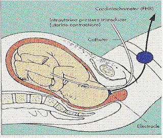
Electronic Fetal Monitoring
- External method involves the use of external transducers placed on the maternal abdomen to assess uterine contractions and the FHR.
- Internal method uses spiral electrode and intrauterine pressure catheter to monitor and record FHR, uterine activity and intrauterine pressure.
External Fetal Monitoring
- FHR: Ultrasound transducer
- High frequency sound waves
- used antepartally and intrapartally
- noninvasive
- Does not require RBOW or dilatation
- Uterine activity: Tocotransducer
- Monitors frequency and duration of contractions by use of a pressure sensing device on abdomen
- Antepartally and intrapartally
- Noninvasive
External Fetal Monitoring

Internal Fetal Monitoring
- FHR: Spiral electrode
- converts fetal ECG to via cardiotachometer
- Used when RBOW
- Cervix dilated
- Penetrates presenting part
- Must be securely attached
- Contractions: IUPC
- measures frequency, duration and intensity of contractions
- two types
- measure intrauterine pressure at catheter tip
- Used with RBOW and dilatation
Internal Fetal Monitoring

Baseline Fetal Heart Rate
- Baseline fetal heart rate
- Tachycardia
- Bradycardia
- Variability
Baseline Fetal Heart Rate
- The average rate during a ten minute segment that excludes periodic and non-periodic (episodic) changes, periods of marked variability, and segments that vary by more than 25 BPM.
- Normal range is 110-160.
Tachycardia
- A baseline FHR above 160 BPM for a ten minute period or greater.
- Can be considered an early sign of fetal hypoxia.
- Can result from maternal or fetal infection, maternal hyperthyroidism, or fetal anemia.
- May occur in response to drugs such as terbutaline, atropine, cocaine.
Bradycardia
- A baseline FHR below 110 BPM for a period greater than 10 minutes.
- Considered a later sign of fetal hypoxia.
- Known to occur before fetal demise.
- Can occur from drugs (anesthetics, prolonged compression of the umbilical cord, maternal hypotension or hypothermia.
Variability
- Described as irregular fluctuations in the baseline FHR of 2 cycles per minute or greater.
- Described as short term or long term.
- Absent or undetected variability
- Minimal variability ( < 5 BPM)
- Moderate variability (6 to 25 BPM)
- Marked variability (> 25 BPM)
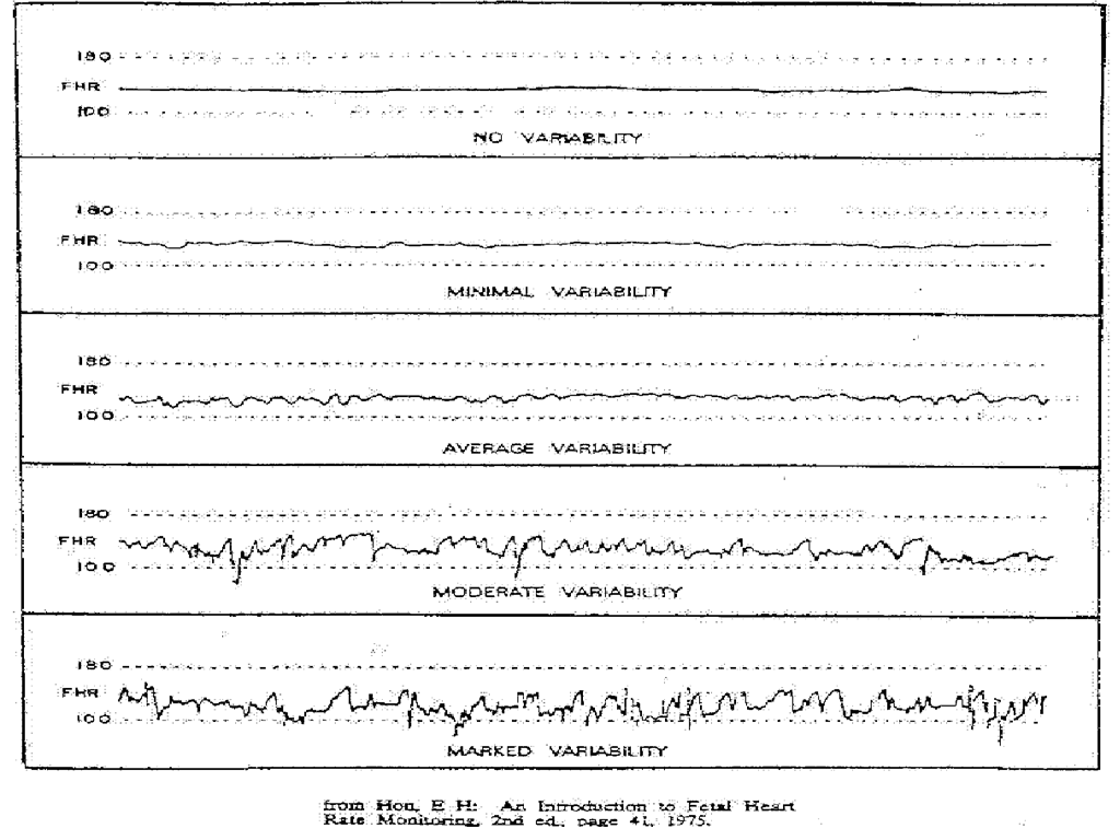
Variability
- In clinical practice used to describe fluctuations in the FHR.
- Absence of variability is considered non-reassuring.
- May result from fetal hypoxemia and acidosis (may be related to drugs).
- A temporary decrease can occur with fetal sleep.
Periodic and Non-periodic FHR Changes
Accelerations
- A visually apparent abrupt increase in FHR above the baseline rate.
- Increase is 15 BPM or greater that lasts 15 seconds or more with return to baseline in less than 2 minutes.
- Can be periodic or non-periodic (episodic).
- Indications of fetal well being.
Decelerations
- May be benign or non-reassuring.
- Described by their relation to the onset and end of the contraction and shape.
- Three types:
- Early decelerations
- Late decelerations
- Variable decelerations
- Prolonged Decelerations
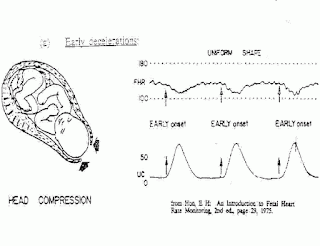
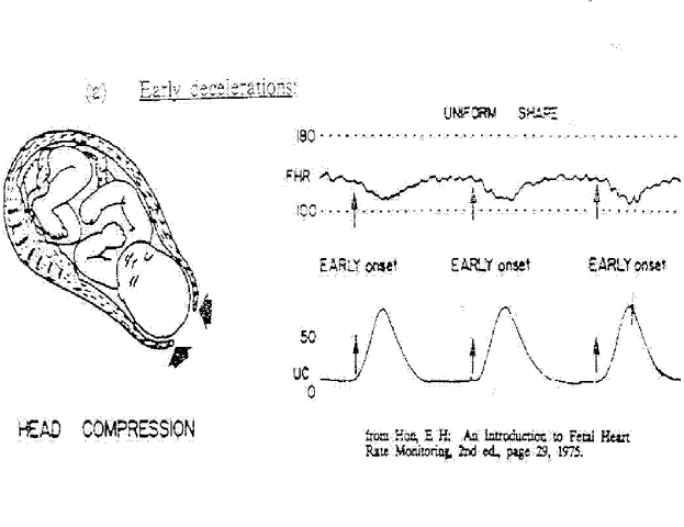
Early Decelerations
- Gradual decrease in and return to FHR baseline.
- In response to head compression.
- Uniform in shape.
- Seen with pushing.
- No intervention required.
Late Decelerations

- Begins after beginning of ctx and ends after end of the contraction.
- May be correctable or ominous
- Caused by uteroplacental insufficiency
Variable Decelerations

- Caused by umbilical cord compression
- Abrupt in descent and return to baseline
- May occur early or late in labor
- May be repetative
Prolonged Decelerations

- May be caused by vaginal exam, spiral electrode application, etc.
- Usually isolated events
- May occur just before fetal death.
Fetal Well-being
- Can be measured by response of the FHR to uterine contractions.
- FHR patterns can be described as reassuring or non-reassuring.
Reassuring FHR patterns
- Baseline FHR in the normal range of 110 to 160 BPM with no periodic changes and a moderate baseline variability.
- Accelerations with fetal movement.
Non-reassuring Patterns
- Progressive increase or decrease in the fetal baseline
- Tachycardia of 160 BPM or more
- Progressive decrease in baseline variability
- Severe variable decelerations
- Late decelerations of any magnitude
- Absence of FHR variability
- Prolonged deceleration
- Severe bradycardia
Normal Uterine Activity
- Occurring every 2 - 5 minutes
- Lasting less than 90 seconds
- Moderate to strong in intensity (by palpation or 100mm Hg by IUPC)
- 30 second lapse period between contractions
- Uterine relaxation between ctx by palpation or 15 mm Hg by IUPC
Fetal Compromise
- The goals of intrapartum FHR monitoring are to identify and differentiate the rassuring from the nonreassuring , which can be indicative of fetal compromise.
- Nonreassuring FHR patterns are those associated with fetal hypoxia (a deficiency in oxygen in the arterial blood) and if uncorrected hypoxia (at the cellular level).
Nonstress Test NST
(pg. 452-454)
(pg. 452-454)
- A reactive NST shows two or more accelerations of 15 bpm or more within 20 minutes of beginning the test.
- A nonreactive NST contains a tracing that does not meet the above criteria. Accelerations are < two in number or < 15 bpm or no accelerations are present.
Contraction Stress Test CST
(pg. 455)
(pg. 455)
- Contractions occurring spontaneously
- Nipple stimulation
- Necessary component is the presence of three uterine contractions of at least 40 sec duration in 10 minute span
- Not done prior to prior to 28 wks gestation
- NEGATIVE, POSITIVE & EQUIVOCAL
Biophysical Profile (BPP)
- Assessment of 5 variables in the fetus that help to evaluate fetal risk: breathing movement, body movement, tone amniotic fluid volume, and fetal heart rate activity.
- A score of 8 to 10 is normal.
- A score of 6 or below indicates fetal compromise
Fetal Acoustic Stimulation Test
Let’s “buzz” the baby!!!!!
Ultrasound
- Most common diagnostic procedure
- 70% of pregnant women have at least one
- Abdominal, vaginal, or labial
- May be basic or limited
- Can evaluate both structural and functional characteristics
- BP diameter, head circumference, femur length, abdominal measurements
- Fetal growth, congenital anomalies, placental growth and location, cervical length

Amniocentesis (pg. 457-459)
- A simple procedure: needle is inserted through the maternal abdomen into the uterine cavity to withdraw a sample of amniotic fluid.
- Early pregnancy: DNA studies
- Late Pregnancy: Lung maturity
- Complications: Preterm labor, fetal scratches, maternal hemorrhage, infection, Rh sensitization (RhoGam may be indicated)
Tocolytic Therapy
- Tocolysis can be achieved by administering drugs that inhibit uterine contractions.
- May be used during management of fetal compromise.
- Magnesium sulfate, terbutaline, nifedipine may be used.
Maternal Positioning
- Maternal supine hypotensive syndrome is caused by the weight and pressure of the gravid uterus on the ascending vena cava when the woman is in a supine position.
- A side-lying position or semi-fowlers position with a lateral tilt to the uterus is recommended.
Other Available Tests
(pg. 459-467)
(pg. 459-467)
- AFP (Amniotic Fluid)
- Rh sensitized pregnancies
- Fetal Maturity
- L/S ratio and PG
- CVS
- Fetoscopy
- Percutaneous Umbilical Blood Sampling
- MRI
EFM: Nursing Diagnosis
- Maternal anxiety related to lack of knowledge about use of electronic fetal monitor.
- Risk for fetal injury related to inaccurate placement of transducers/electrodes, misinterpretation of results or failure to use other assessment techniques to monitor fetal well-being.
Nursing Assessment & Diagnosis
- Knowledge Deficit related to insufficient information about the fetal assessment test and its purpose, benefits, risks, and alternatives
- Fear related to the specific test or possible unfavorable results
- Disruption in bonding due to high risk label
Questions?
Source : Abraham Baldwin Agricultural College
About: ABAC Enrollment Fall 20113,248 students from 17 states, 22 countries, and 149 Georgia counties
No comments:
Post a Comment