Introduction and Tissues
What is Anatomy?
Each of these build upon one another to make up the next level:
Anatomical Directions
Tissues: groups of cells closely associated that have a similar structure and perform a related function


Defense from Infection

Image
Anatomy of a Long Bone


Muscle Tissue
(each skeletal muscle is an organ)

Nervous Tissue
Human Anatomy
BIOL 1010
Liston Campus

Liston Campus
What is Anatomy?
- Anatomy (= morphology): study of body’s structure
- Physiology: study of body’s function
- Structure reflects Function!!!
- Branches of Anatomy
- Gross: Large structures
- Surface: Landmarks
- Histology: Cells and Tissues
- Developmental: Structures change through life
- Embryology: Structures form and develop before birth
Each of these build upon one another to make up the next level:
- Chemical level
- Cellular
- Tissue
- Organ
- Organ system
- Organism
- Chemical level
- Atoms combine to make molecules
- 4 macromolecules in the body
- Carbohydrates
- Lipids
- Proteins
- Nucleic acids
Hierarchy of Structural Organization
- Cellular
- Made up of cells and cellular organelles (molecules)
- Cells can be eukaryotic or prokaryotic
- Organelles are structures within cells that perform dedicated functions (“small organs”)
- Tissue
- Collection of cells that work together to perform a specialized function
- 4 basic types of tissue in the human body:
- Epithelium
- Connective tissue
- Muscle tissue
- Nervous tissue
- Organ
- Made up of tissue
- Heart
- Brain
- Liver
- Pancreas, etc……
- Organ system (11)
- Made up of a group of related organs that work together
- Integumentary
- Skeletal
- Muscular
- Nervous
- Endocrine
- Cardiovascular
- Lymphatic
- Respiratory
- Digestive
- Urinary
- Reproductive
Hierarchy of Structural Organization
- Organism
- An individual human, animal, plant, etc……
- Made up all of the organ systems
- Work together to sustain life
Anatomical Directions
- Anatomical position
- Regions
- Axial vs. Appendicular
- Anatomical Directions-It’s all Relative!
- Anterior (ventral) vs. Posterior (dorsal)
- Medial vs. Lateral
- Superior (cranial) vs. Inferior (caudal)
- Superficial vs. Deep
- Proximal vs. Distal
- Anatomical Planes
- Frontal = Coronal
- Transverse = Horizontal = Cross Section
- Sagittal
| Anterior – (ventral) Closer to the front surface of the body |
Posterior – (dorsal) Closer to the rear surface of the body |
Frontal Plane |
| Medial – Lying closer to the midline |
Lateral – Lying further away from the midline |
Sagittal Plane |
| Superior – (cranial) Closer to the head in relation to the entire body (More General) |
Inferior – (caudal) Away from the head or towards the lower part of the body |
Horizontal Plane |
| Superficial – Towards the surface |
Deep – Away from the surface |
Surface of body or organ |
| Proximal – Closer to the origin of a body part (More Specific)  |
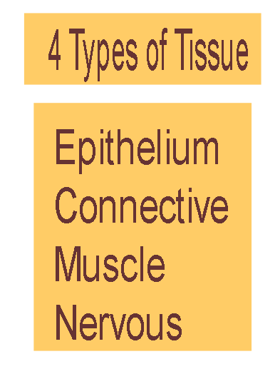
- Four types of tissue
- Epithelial = covering/lining
- Connective = support
- Muscle = movement
- Nervous = control
- Most organs contain all 4 types
- Tissue has non-living extracellular material between its cells
- Functions
- Protection
- Secretion
- Absorption
- Ion Transport
- Cellularity
- Composed of cells
- Specialized contacts
- Joined by cell junctions
- Polarity
- Apical vs. Basal surfaces differ
- Supported by connective tissue
- Avascular
- Innervated
- Highly regenerative
- Layers
- Simple
- Stratified
- Stratified layers characterized by shape of apical layer
- Psuedostratified
- Shapes
- Squamous
- Cuboidal
- Columnar
- Transitional

Types of Epithelium
- Simple squamous (1 layer)
- Lungs, blood vessels, ventral body cavity
- Simple cuboidal
- Kidney tubules, glands
- Simple columnar
- Stomach, intestines
- Pseudostratified columnar
- Respiratory passages (ciliated version)
- Stratified squamous (>1 layer)
- Epidermis, mouth, esophagus, vagina
- Named so according to apical cell shape
- Regenerate from below
- Deep layers cuboidal and columnar
- Transitional (not shown)
- Thins when stretches
- Hollow urinary organs
- Endothelium
- Simple squamous epithelium that lines vessels
- e.g. lymphatic & blood vessel
- Mesothelium
- Simple squamous epithelium that forms the lining of body cavities
- e.g. pleura, pericardium, peritoneum
- Microvilli: (ex) in small intestine
- Finger-like extensions of the plasma membrane of apical epithelial cell
- Increase surface area for absorption
- Cilia: (ex) respiratory tubes
- Whip-like, motile extension of plasma membrane
- Moves mucus, etc. over epithelial surface 1-way
- Cells are connected to neighboring cells via:
- Contour of cells-wavy contour fits together
- Cell Junctions (3 common)
- Desmosomes
- Proteins hold cells together to maintain integrity of tissue
- Tight Junctions
- Plasma membrane of adjacent cells fuse, nothing passes
- Gap junction
- Proteins allow small molecules to pass through
Features of the Basal Surface of Epithelium
- Basement membrane
- Sheet between the epithelial and connective tissue layers
- Attaches epithelium to connective tissue below
- Made up of:
- Basal lamina: thin, non-cellular, supportive sheet made of proteins
- Superficial layer
- Acts as a selective filter
- Assists epithelial cell regeneration by moving new cells
- Reticular fiber layer
- Deeper layer
- Support
- Epithelial cells that make and secrete a product
- Products are water-based and usually contain proteins
- Classified as:
- Unicellular vs. multicellular
- Exocrine vs. Endocrine
- Exocrine Glands
- Secrete substance onto body surface or into body cavity
- Activity is local
- Have ducts
- Unicellular or Multicellular
- (ex) goblet cells, salivary, mammary, pancreas, liver
- Endocrine Glands
- Secrete product into blood stream
- Either stored in secretory cells or in follicle surrounded by secretory cells
- Hormones travel to target organ to increase response (excitatory)
- No ducts
- (ex) pancreas, adrenal, pituitary, thyroid


Connective Tissue (CT):
most abundant and diverse tissue
most abundant and diverse tissue
- Four Classes
- Functions include connecting, storing & carrying nutrients, protection, fight infection
- CT contains large amounts of non-living extracellular matrix
- Contains a variety of cells and fibers
- Some types vascularized
- All CT originates from mesenchyme
- Embryonic connective tissue
- Fibers For Support
- Reticular:
- form networks for structure & support
- (ex) cover capillaries
- Collagen:
- strongest, most numerous, provide tensile strength
- (ex) dominant fiber in ligaments
- Elastic:
- long + thin, stretch and retain shape
- (ex) dominant fiber in elastic cartilage
Components of Connective Tissue
- Fibroblasts:
- cells that produce all fibers in CT
- produce + secrete protein subunits to make them
- produce ground matrix
- Interstitial (Tissue) Fluid
- derived from blood in CT proper
- medium for nutrients, waste + oxygen to travel to cells
- found in ground matrix
- Ground Matrix (substance):
- part of extra-cellular material that holds and absorbs interstitial fluid
- Made and secreted by fibroblasts
- jelly-like with sugar & protein molecules
- Connective Tissue Proper
Two kinds: Loose CT & Dense CT
- Functions
- Support and bind to other tissue
- Hold body fluids
- Defends against infection
- Stores nutrients as fat
- Each function performed by different kind of fibers and cells in specific tissue
Defense from Infection
- Areolar tissue below epithelium is body’s first defense
- Cells travel to CT in blood
- Macrophages-eat foreign particles
- Plasma cells-secrete antibodies, mark molecules for destruction
- Mast cells-contain chemical mediators for inflammation response
- White Blood Cells = neutrophils, lymphocytes, eosinophils-fight infection
- Ground substance + cell fibers-slow invading microorganisms
- Areolar CT
- All types of fibers present
- All typical cell types present
- Surrounds blood vessels and nerves
Specialized Loose CT Proper
- Adipose tissue
- Loaded with adipocytes, highly vascularized, high metabolic activity
- Insulates, produces energy, supports
- Found in hypodermis under skin
- Reticular CT
- Contains only reticular fibers
- Forms caverns to hold free cells, forms internal “skeleton” of some organs
- Found in bone marrow, holds blood cells, lymph nodes, spleen
- Contains more collagen
- Can resist extremely strong pulling forces
- Regular vs. Irregular
- Regular-fibers run same direction, parallel to pull
- (eg) fascia, tendons, ligaments
- Irregular-fibers thicker, run in different directions
- (eg) dermis, fibrous capsules at ends of bones

- Chondroblasts produce cartilage
- Cartilage
- Chondrocytes mature cartilage cells
- Reside in lacunae
- More abundant in embryo than adult
- Firm, Flexible
- Resists compression
- (eg) trachea, meniscus
- Avascular (chondrocytes can function w/ low oxygen)
- NOT Innervated
- Perichondrium
- dense, irregular connective tissue around cartilage
- growth/repair of cartilage
- resists expansion during compression of cartilage
Cartilage in the Body
- Three types:
- Hyaline
- most abundant
- fibers in matrix
- support via flexibility/resilience
- (eg) at limb joints, ribs, nose
- Elastic
- many elastic fibers in matrix too
- great flexibility
- (eg) external ear, epiglottis
- Fibrocartilage
- resists both compression and tension
- (eg) meniscus, annulus fibrosus
- Well-vascularized
- Function:
- support (eg) pelvic bowl, legs
- protect (eg) skull, vertebrae
- mineral storage (eg) calcium, phosphate (inorganic component)
- movement (eg) walk, grasp objects
- blood-cell formation (eg) red bone marrow
- Osteoblasts
- Secrete organic part of bone matrix
- Osteocytes
- Mature bone cells
- Sit in lacunae
- Maintain bone matrix
- Osteoclasts
- Degrade and reabsorb bone
- Periosteum
- Bone Tissue: (a bone is an organ)
- External layer of CT that surrounds bone
- Outer: Dense irregular CT
- Inner: Osteoblasts, osteoclasts
- Endosteum
- Internal layer of CT that lines cavities and covers trabeculae
- Contains osteoblasts and osteoclasts
- External layer
- Osteon (Haversian system)
- Parallel to the long axis of the bone
- Groups of concentric tubules (lamella)
- Lamella = layer of bone matrix where all fibers run in the same direction
- Adjacent lamella fibers run in opposite directions
- Haversian Canal runs through center of osteon
- Contains blood vessels and nerves
- Connected to each other by perforating (Volkman) canals
- Interstitial lamellae fills spaces and forms periphery
- Spongy bone (cancellous bone): internal layer
- Trabeculae: small, needle-like pieces of bone form honeycomb
- each made of several layers of lamellae + osteocytes
- no canal for vessels
- space filled with bone marrow
- not as dense, no direct stress at bone’s center
Image
Anatomy of a Long Bone
- Diaphysis
- Medullary Cavity
- Nutrient Artery & Vein
- 2 Epiphyses
- Epiphyseal Plates
- Epiphyseal Artery & Vein
- Periosteum
- Does not cover epiphyses
- Endosteum
- Covers trabeculae of spongy bone
- Lines medullary cavity of long bones
- Intramembranous Ossification
- Membrane bones: most skull bones and clavicle
- Osteoblasts in membrane secrete osteoid that mineralizes
- Endochondral Ossification: All other bones
- Begins with a cartilaginous model
- Cartilage calcifies
- Medullary cavity is formed by action of osteoclasts
- Epiphyses grow and eventually calcify
- Epiphyseal plates remain cartilage for up to 20 years
- Appositional Growth = widening of bone
- Bone tissue added on surface by osteoblasts of periosteum
- Medullary cavity maintained by osteoclasts
- Lengthening of Bone
- Epiphyseal plates enlarge by chondroblasts
- Matrix calcifies (chondrocytes die and disintegrate)
- Bone tissue replaces cartilage on diaphysis side
- REMODELING
- Due to mechanical stresses on bones, their tissue needs to be replaced
- Osteoclasts-take up bone ( = breakdown) release Ca 2++ , PO 4 to body fluids from bone
- Osteoblasts-form new bone by secreting osteoid
- Ideally osteoclasts & osteoblasts work at the sam
- GROWTH
- e rate!
- Components of Bone Tissue Summarized
- Blood: Atypical Connective Tissue
- Function:
- Transports waste, gases, nutrients, hormones through cardiovascular system
- Helps regulate body temperature
- Protects body by fighting infection
- Derived from mesenchyme
- Hematopoiesis: production of blood cells
- Occurs in red bone marrow
- In adults, axial skeleton, girdles, proximal epiphyses of humerus and femur
Blood Cells
- Erythrocytes: (RBC) small, oxygen-transporting
- most abundant in blood
- no organelles, filled w/hemoglobin
- pick up O 2 at lungs, transport to rest of body
- Leukocytes: (WBC) complete cells , 5 types
- fight against infectious microorganisms
- stored in bone marrow for emergencies
- *Platelets = Thrombocytes:
- fragments of cytoplasm
- plug small tears in vessel walls, initiates clotting
- Components of Blood Summarized

Muscle Tissue
- Muscle cells/fibers
- Elongated
- Contain many myofilaments: Actin & Myosin
- FUNCTION
- Movement
- Maintenance of posture
- Joint Stabilization
- Heat Generation
- Three types: Skeletal, Cardiac, Smooth
(each skeletal muscle is an organ)
- Cells
- Long and cylindrical, in bundles
- Multinucleate
- Obvious Striations
- Skeletal Muscles-Voluntary
- Connective Tissue Components:
- Endomysium-surrounds fibers
- Perimysium-surrounds bundles
- Epimysium-surrounds the muscle
- Attached to bones, fascia, skin
- Origin & Insertion
- Cells
- Branching, chains of cells
- Single or Binucleated
- Striations
- Connected by Intercalated discs
- Cardiac Muscle-Involuntary
- Myocardium-heart muscle
- Pumps blood through vessels
- Connective Tissue Component
- Endomysium: surrounding cells
- Cells
- Single cells, uninucleate
- No striations
- Smooth Muscle-Involuntary
- 2 layers-opposite orientation (peristalsis)
- Found in hollow organs, blood vessels
- Connective Tissue Component
- Endomysium: surrounds cells

Nervous Tissue
- Neurons: specialized nerve cells conduct impulses
- Cell body, dendrite, axon
- Characterized by:
- No mitosis (cell replication)
- Longevity
- High metabolic rate
- Support cells (= Neuroglial): nourishment, insulation, protection
- Satellite cells-surround cell bodies within ganglia
- Schwann cells-surround axons (PNS)
- Microglia-phagocytes
- Oligodendrocytes-produce myelin sheaths around axons
- Ependymal cells-line brain/spinal cord, ciliated, help circulate CSF
- Brain, spinal cord, nerves
- Functions
- Protection
- Mechanical, thermal, chemical, UV
- Cushions & insulates deeper organs
- Prevention of water loss
- Thermoregulation
- Excretion
- Salts, urea, wa
- Sensory reception
Microanatomy - Layers of the Skin
- Epidermis
- Epithelium
- Dermis
- Connective tissue
- Hypodermis / subcutis
- Loose connective tissue
- Anchors skin to bone or muscle
- Skin Appendages = outgrowths of epidermis
- Hair follicles
- Sweat and Sebaceous glands
- Nails
Cell Layers of the Epidermis
- Stratum corneum
- Dead keratinocytes
- Stratum lucidum
- Only in “thick” skin
- Dead keratinocytes
- Stratum granulosum
- Water proofing
- Stratum spinosum
- Resists tears and tension
- Stratum basale
- Sensory receptors
- Melanocytes
- Keratinocytes (in all layers)
- Highly innervated
- Highly vascularized
- Collagen & Elastic fibers
- 2 layers:
- Papillary layer (20%)
- Areolar CT
- Collagen & Elastic fibers
- Innervation
- Hair follicles
- Reticular layer (80%)
- Dense irregular CT
- Glands
- sebum
- 2.5 million sweat glands!!
- Smooth muscle fibers
- Innervation
- Also called superficial fascia
- Areolar & Adipose Connective Tissue
- Functions
- Store fat
- Anchor skin to muscle, etc.
- Insulation
LUMEN
- Serosa – suspends organ in the peritoneal
- Tunica Mucosa
- Lamina epithelialis
- Lamina propria
- Lamina muscularis mucosa
- Tunica Submucosa
- Tunica Muscularis
- Inner circular
- Outer longitudinal
- Tunica Adventitia / Serosa
- Adventitia – covers organ
- cavity
